Your Glaucoma Suspect Visit at a Glance
A suspect label is a reason to look closer
Being called a glaucoma suspect does not mean you have confirmed glaucoma. Some people receive the label because eye pressure is higher than usual without nerve damage, while others have a suspicious optic nerve even when pressure is within a usual range. 1 The visit should tell you what raised concern, what the tests show now, and what happens next.
The evaluation builds a starting point
Most glaucoma suspects have no symptoms, so feeling or seeing normally does not replace monitoring. 1 Evaluation may combine eye-pressure measurement, a visual-field test, optic-nerve examination or imaging, drainage-angle assessment, and corneal-thickness measurement. Some tests are repeated only when clinically indicated. 2
Why Several Glaucoma Tests Are Used Together
One pressure reading does not settle the diagnosis
A person can be a glaucoma suspect with high pressure and no visible damage, or with a suspicious optic nerve despite pressure in a usual range. 1 Glaucoma assessment therefore considers pressure alongside the optic nerve, visual field, drainage angle, and other findings rather than relying on one number. 2 Ask which finding led to your referral.
Structure and vision answer different questions
Optic-nerve examination and OCT imaging assess the eye’s structure, while standard automated perimetry tests the visual field. The clinician uses these different results together rather than asking one test to answer every question. 2 A result can be reassuring in one area and uncertain in another.
Baseline records make later comparison possible
NICE recommends an optic-nerve image at diagnosis for baseline documentation and repeat nerve or field assessment when clinically indicated. A baseline gives the care team something specific to compare with future results; it is not a prediction that vision will worsen. 2
What to Bring and Plan Before the Visit
Gather prior eye records if they are available
Ask the referring clinic to send earlier pressure readings, visual fields, optic-nerve photographs, OCT scans, and visit notes. Bring copies if you have them. Write down which eye raised concern and why. Do not delay the evaluation if old records cannot be found; give the clinic the details needed to request them.
List health history, medicines, and family history
Bring a list of prescription and nonprescription medicines, vitamins, and eye drops. Include how often you use them. Long-term steroid medicine, prior eye injury, thin central corneas, high eye pressure, and a family history of glaucoma are among factors associated with glaucoma risk. 1 Tell the clinician about previous eye surgery and close relatives with glaucoma.
Bring the glasses and aids you normally use
Bring your current distance and reading glasses. If you use hearing aids, mobility aids, or an interpreter, arrange them as you usually would for a medical visit. Tell the clinic before testing if you have difficulty positioning at a machine or pressing a response button. For a related symptom pattern, read Caffeine With Glaucoma and What the Evidence Means.
Plan for possible dilation
After a dilated eye examination, vision may be blurry and the eyes may be sensitive to light for a few hours. The National Eye Institute recommends asking a friend or family member to drive you home and bringing sunglasses. 3 Ask the clinic in advance whether dilation is likely and what transport plan it recommends.
What May Happen During the Evaluation
Visual acuity and pupil checks may come first
A dilated eye examination may include tests of visual clarity, side vision, eye movements, pupil responses, and eye pressure. 3 The order varies by clinic. Tell the technician when an instruction is unclear rather than guessing what to do.
Tonometry measures pressure inside the eye
Tonometry measures eye pressure. The test may use a quick puff of air or a device that gently touches the eye, and the National Eye Institute describes it as not painful. A pressure reading is one part of the evaluation. 3 2 Ask which method was used if you are comparing it with an older record.
A visual-field test checks side vision
Standard automated perimetry is used to assess the visual field and may be repeated when needed to establish or monitor a finding. 2 Follow the test instructions and tell the technician if you lose focus or need a pause. An unfamiliar test does not require a perfect performance.
Dilation and imaging examine the optic nerve
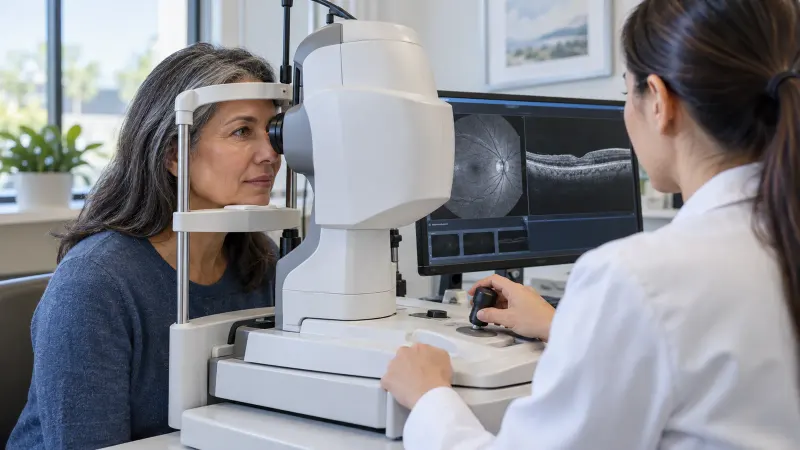
Dilating drops widen the pupil so the doctor can examine the inside of the eye. Optic-nerve photographs or OCT may be used as baseline images for later comparison. The clinician may use both direct examination and imaging because they provide different records of the nerve. 3 2
Gonioscopy and pachymetry add other details
A glaucoma-suspect evaluation may include gonioscopy to assess the drainage angle and pachymetry to measure central corneal thickness. 2 4 Ask what each test is meant to clarify in your case and whether it needs to be repeated later.
How to Understand the Results
If no glaucoma damage is found
Some glaucoma suspects have higher pressure without signs of optic-nerve damage, and some have a suspicious nerve appearance without confirmed glaucoma. A reassuring visit can still end with monitoring for change. 1 Ask which result is normal, which finding remains suspicious, and what will be compared next time.
If the pressure is higher than expected
Higher eye pressure can lead to glaucoma-suspect status, but pressure by itself does not show whether the optic nerve is damaged. Corneal-thickness, optic-nerve, drainage-angle, and visual-field findings add context to the pressure result. 1 2 Ask whether the term “ocular hypertension” applies.
If the visual field is uncertain
Visual-field testing may be repeated when clinically indicated, and the same measurement strategy is used when a prior defect has been detected. 2 Ask whether the result showed a consistent pattern, was limited by test reliability, or needs confirmation. An uncertain test should be recorded as uncertain rather than described as normal.
If OCT or the optic nerve looks suspicious
OCT, optic-nerve imaging, and careful nerve examination provide complementary information for glaucoma suspects. 4 Ask which part of the nerve or retinal nerve-fiber layer raised concern and whether an older image is available for comparison.
If treatment is discussed
An ophthalmologist may monitor a glaucoma suspect for change over time and begin treatment if it becomes necessary. Treatment decisions follow the combined findings and risk assessment rather than the suspect label alone. 1 2 Ask what risk the treatment is intended to lower, what result would show it is working, and what alternatives were considered.
What the Tests Can and Cannot Tell You
A normal-feeling eye can still need follow-up
Most glaucoma suspects have no symptoms, and an ophthalmologist monitors for changes over time. 1 This can feel frustrating when vision seems normal. The practical answer is to leave with a specific next date and a clear reason for repeating each test.
A single visit may leave some uncertainty
NICE recommends repeat visual-field, drainage-angle, or optic-nerve assessment when clinically indicated and uses current risk to set the next assessment time. An uncertain result may need confirmation and does not by itself establish glaucoma. 2
Most test effects are temporary
Eye-pressure testing is described as not painful, while dilation may cause temporary blur and light sensitivity for a few hours. 3 Tell the team if positioning, light, or concentration makes a test difficult so that context is recorded with the result.
When to Keep the Plan or Seek Faster Care
Keep the scheduled suspect follow-up
Glaucoma suspects often have no symptoms, so monitoring is used to check for change over time. Keep the planned return even when everyday vision feels unchanged. 1 Contact the clinic if you cannot attend so a new date and test plan are documented.
Get urgent care for sudden painful vision loss
Sudden painful vision loss can be acute angle closure, which is an ophthalmic emergency and can have a good prognosis when treated promptly. Seek emergency eye care now for this sudden pattern rather than waiting for a routine glaucoma-suspect appointment. 5 An angle-closure attack may also cause a red eye, blurred vision, halos, headache, nausea, or vomiting. 1
Call sooner when the clinic tells you a test needs repeating
If a result was unreliable, incomplete, or concerning, ask who will arrange the repeat test and by what date. Call the clinic if that appointment does not appear. Keep copies of your summary so another clinician can see what is known and what remains uncertain. You can compare this topic with Setting a Target Eye Pressure in Open-Angle Glaucoma.
Questions About the Glaucoma Suspect Label
Does glaucoma suspect mean I have glaucoma?
No. Some people are glaucoma suspects because pressure is higher than usual without nerve damage. Others have a suspicious optic nerve even when pressure is in a usual range. The evaluation looks for evidence of damage and establishes what should be monitored. 1 Ask the clinician which finding led to the label in your record.
Can I have glaucoma with a normal pressure reading?
Yes. Normal-tension glaucoma can occur when pressure readings are within usual ranges but visual-field or optic-nerve damage is present. This is why the visit combines pressure with nerve examination, imaging, and a visual-field test. 1 2 Ask how your pressure fits the other results rather than interpreting one number on your own.
Does high eye pressure always mean nerve damage?
No. Ocular hypertension means pressure is higher than usual without signs of damage, and it can lead to glaucoma-suspect status. The clinician considers pressure with the optic nerve, cornea, drainage angle, visual field, and other risk factors before recommending monitoring or treatment. 1 2 Ask which risk factor carries the most weight in your plan.
Will I need glaucoma drops after the evaluation?
Not necessarily. An ophthalmologist may monitor a glaucoma suspect and begin treatment only if it is needed. 1 Ask what current finding would support treatment, what change would trigger treatment later, and how the team would judge whether it is helping. Do not start or stop drops based on a general article.
Why might the visual-field test be repeated?
Visual-field testing is repeated when clinically indicated to confirm or monitor a finding, and the same testing strategy is used when a defect has already been detected. 2 Ask whether the first result was reliable, whether the pattern matches the optic nerve, and when a repeat would be most useful.
Questions About Tests and Follow-Up
What does OCT add to the examination?
OCT is one form of optic-nerve imaging used in glaucoma-suspect evaluation, and baseline nerve imaging can be compared with later results. 4 2 Ask what structure was measured, whether the scan quality was acceptable, and which future scan should be compared with it. Keep the report for future visits.
Why would I need gonioscopy or pachymetry?
Gonioscopy assesses the drainage angle, while pachymetry measures central corneal thickness. Both can be part of a glaucoma-suspect evaluation. 2 4 Ask whether either result changed how the pressure, risk, or next appointment was interpreted. Ask for each result in plain language, and keep the measurements with your visit summary.
Can I drive after my pupils are dilated?
Dilation can cause blurry vision and light sensitivity for a few hours, and the National Eye Institute recommends asking a friend or family member to drive you home. 3 Ask the clinic about its instructions before the appointment, arrange transport if needed, and bring sunglasses for comfort. Plan this before the drops are placed.
How often will I need to return?
The next assessment time should be based on the current risk of developing glaucoma and the risk of sight loss, so there is no single interval that fits every suspect. 2 Ask for a specific return date, which tests will be repeated, and who will contact you if the plan changes.
Questions to Ask Your Doctor
- What finding made me a glaucoma suspect?
- Do the optic nerve, visual field, pressure, angle, and corneal thickness tell a consistent story?
- Which result is the baseline for future comparison?
- Was any test unreliable or incomplete?
- What would change the plan from monitoring to treatment?
- When is my next assessment, and who will arrange it?
Sources
- American Academy of Ophthalmology (2026). Understanding Glaucoma: Symptoms, Causes, Diagnosis, Treatment.
- National Institute for Health and Care Excellence (2025). Glaucoma: diagnosis and management - Recommendations.
- National Eye Institute (2025). Get a Dilated Eye Exam.
- American Academy of Ophthalmology EyeWiki (2026). Glaucoma Screening.
- BMJ Clinical Evidence (2015). Glaucoma: acute and chronic primary angle-closure.