Corneal cross-linking for keratoconus is designed to slow or stop the cornea from becoming thinner and steeper. The procedure strengthens links within corneal tissue using riboflavin and controlled ultraviolet light. Its main purpose is stability. You may still need glasses or specialty contact lenses afterward. For a related symptom pattern, read How to Compare LASIK and PRK for Refractive Surgery.
At a Glance
The treatment goal
- Cross-linking targets documented keratoconus progression.
- It aims to preserve usable vision by stabilizing corneal shape.
- It does not remove keratoconus or promise sharper vision.
- Follow-up remains necessary after the cornea heals.
Progression matters more than the label alone
Some people have a stable corneal shape for years, while others change over a shorter period. A cornea specialist compares maps, thickness measurements, prescription changes, and vision over time. The American Academy of Ophthalmology EyeWiki review of keratoconus describes monitoring as the step that helps determine when cross-linking is indicated.
What keratoconus changes in the eye
The cornea loses its regular curve
The cornea is the clear front surface of the eye. Keratoconus causes it to thin and bulge into a less regular shape. Light then scatters instead of focusing cleanly, which can cause blurred vision, ghost images, glare, and astigmatism that changes more than expected. You can compare this topic with What Helps Eye Strain From Screens and What Does Not.
Glasses may stop giving crisp vision
Early changes may respond to a new glasses prescription. As the surface becomes more irregular, glasses may not correct all the blur. Rigid or scleral lenses can create a smoother optical surface, but they improve focus rather than stop the underlying shape change.
Eye rubbing can add stress
Frequent forceful rubbing is associated with keratoconus progression. Treating allergy or irritation can make it easier to stop. Pat or use a cool compress instead of pressing on the eyes, and tell the specialist about itching, sleep position, or habits that put pressure on one eye.
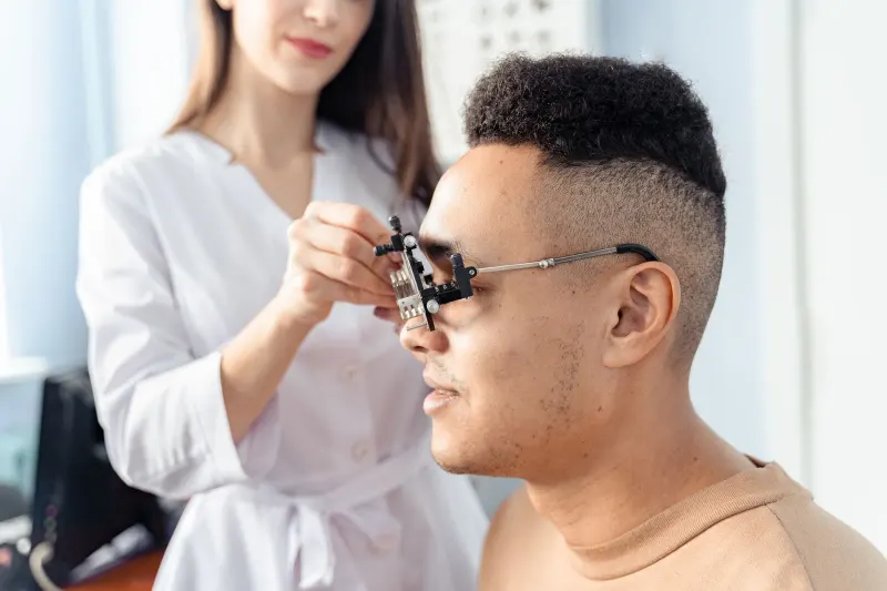
How doctors confirm progression
Corneal maps show shape over time
Topography and tomography create detailed maps of the front and back corneal surfaces. The specialist compares scans rather than relying on one color map. Small differences can come from dry eye, contact lens wear, or measurement quality, so repeatable change matters.
Thickness and prescription add context
The doctor also measures corneal thickness and reviews astigmatism, best-corrected vision, and lens fit. A rising prescription alone does not prove progression. The full pattern helps separate true structural change from an unstable tear film or an inaccurate scan.
Age affects the monitoring plan
Keratoconus can progress faster in children and younger adults, so specialists may monitor them more closely. Older age does not guarantee stability. The schedule should reflect prior scans, symptoms, eye rubbing, family history, and how quickly vision has changed.
What happens during corneal cross-linking
Riboflavin and light strengthen tissue
The clinician places riboflavin drops on the cornea and applies a controlled ultraviolet light. Some methods remove the thin surface layer of the cornea first, while other methods leave it in place. The approved approach, corneal thickness, and specialist's judgment determine the plan.
The surface needs time to heal
With surface-removal treatment, the clinic may place a bandage contact lens while the epithelium grows back. Discomfort, light sensitivity, tearing, and hazy vision are common early in recovery. Follow the prescribed drop and visit schedule, and do not rub the eye.
Vision can fluctuate before it settles
The cornea may remain blurry or uneven while it heals. A temporary change does not show the final result. The specialist may delay a new glasses or contact lens fitting until measurements become stable enough to guide correction.
What corneal cross-linking for keratoconus can and cannot do
Stability is the main outcome
The EyeWiki clinical overview of corneal cross-linking identifies prevention of ectasia progression as the main purpose. Some people gain lines of corrected vision or have a flatter cornea, but those changes are not guaranteed and should not be the reason for promising spectacle-free vision.
Vision correction may still be needed
- Glasses may work when irregularity remains mild.
- Rigid or scleral lenses may provide clearer vision for an irregular cornea.
- A new fitting may be needed after the cornea stabilizes.
- Further surgery is reserved for selected cases with major scarring or poor function.
Cross-linking has risks and limits
Possible problems include infection, haze, scarring, delayed surface healing, and failure to stop progression. A thin cornea or active surface disease may change eligibility. Ask which technique is planned and why it fits your measurements.
When to call during recovery
Worsening symptoms need prompt review
Contact the treating clinic for increasing pain, new discharge, worsening redness, a sudden drop in vision, or a bandage lens that falls out. Do not replace the lens yourself unless the clinician gave specific instructions.
Keep every follow-up scan
Healing checks protect the cornea early, and later scans show whether the shape has stabilized. Even after treatment, report new ghosting or prescription changes. People comparing lens-based vision options can also review who scleral contact lenses may help.
Plan around temporary visual limits
Arrange transport for the procedure and ask when work, school, screens, exercise, and driving can resume. The treated eye may be light-sensitive or blurred early on, so the clinic's restrictions matter more than a fixed online recovery calendar. Tell the team if your other eye also has limited vision because that can change practical support needs.
Common Questions About Corneal Cross-Linking
Does cross-linking make keratoconus go away?
No. It aims to stabilize the cornea. It does not restore a normal corneal shape or remove the need for vision correction.
How does a doctor know the condition is progressing?
The doctor compares corneal maps, thickness, prescription, and corrected vision across visits. Repeatable change carries more weight than one scan.
Can both eyes need treatment?
Keratoconus can affect each eye differently. A specialist evaluates and schedules each eye based on its own progression and safety measurements.
Can I wear contacts after cross-linking?
Many people return to contact lenses after healing. The timing and need for refitting depend on surface recovery and stable measurements.