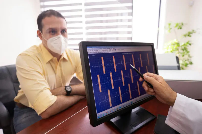
The most important test for suspected conversion from dry to wet age-related macular degeneration is often structural optical coherence tomography, or OCT, interpreted with symptoms and a dilated retinal examination. OCT can show fluid or other structural changes associated with active macular neovascularization. OCT angiography and dye-based angiography may be added when the diagnosis or activity is unclear.
No home check or single office image can guarantee that conversion has not occurred. New distortion, a dark or blank central area, or a sudden decline in central vision should prompt timely contact with the eye clinic, even if the last scan was reassuring. For a related symptom pattern, read What Retinal OCT Biomarkers Can Show.
Dry to wet AMD conversion means age-related macular degeneration has developed abnormal leaky blood vessels or fluid under or within the retina. Catching this change matters because wet AMD can affect central vision quickly.
Patients with dry AMD may be monitored with dilated exams, retinal photos, optical coherence tomography, and home symptom checks. The goal is to identify treatable changes promptly, not to make patients anxious about every fluctuation.
What matters first for tests for dry to wet AMD conversion?
Dry AMD can remain stable, but some eyes develop wet AMD. The weight of that detail rests on how well it fits the history, measurements, and care timeline.
- OCT can show fluid or structural change before symptoms are obvious.
- New distortion, a blank spot, or sudden central blur should be reported promptly.
- Testing cannot prevent conversion, but it can help catch changes sooner.
Why Dry To Wet Amd Conversion Matters
Dry AMD can remain stable for long periods, but some eyes convert to wet AMD. Wet AMD is not diagnosed by symptoms alone because early fluid can appear before vision changes are obvious.
Optical coherence tomography is especially useful because it shows cross-sectional retinal detail. It can reveal fluid or structural change that may not be visible to the patient in daily life.
The National Eye Institute describes wet AMD as a form involving abnormal blood vessels that can leak fluid or blood. That is why retinal imaging and symptom awareness are both important.
What does retinal monitoring measure?
The purpose of retinal monitoring is to compare new fluid, bleeding, or retinal thickening on imaging with change in central vision, reading, or face recognition. That retinal monitoring comparison helps the clinician decide whether the result answers the tests for dry to wet AMD conversion question or needs confirmation from another part of the examination.
Interpret retinal monitoring alongside new distortion on lines or grids and differences between one eye and the other. When the retinal monitoring findings conflict, review acquisition quality and consider a repeat or different method before accepting the first output.
Describe the daily task connected with new fluid, bleeding, or retinal thickening on imaging, and note whether the change is stable, intermittent, or worsening. That history gives retinal monitoring a functional context without asking the measurement to diagnose more than it can show.
- New fluid, bleeding, or retinal thickening on imaging
- Change in central vision, reading, or face recognition
- New distortion on lines or grids
- Differences between one eye and the other
What can retinal monitoring results establish?
A normal home grid or good day of reading does not replace scheduled retinal care. Symptoms can be subtle, and one eye may compensate for the other.
Testing also helps avoid assuming that every blur is wet AMD. Cataract, dry eye, glasses changes, and other retinal problems can affect vision too.
A scan may show findings that need monitoring but not immediate treatment. Patients should ask what was seen, what changed, and what symptom would make the timeline faster.
- Ask what the result means for your specific diagnosis.
- Ask whether the finding is new, stable, or uncertain.
- Ask whether repeat testing or imaging is recommended.
- Ask what symptoms should prompt faster contact before the next visit.
What happens during retinal monitoring?
The visit may include a dilated retinal exam, OCT, fundus photos, and sometimes dye-based or noninvasive vascular imaging. Each test provides a different view of the macula.
The clinician may compare the scan with prior images. This comparison is often more useful than looking at one image alone because AMD care depends on change over time.
If wet AMD is suspected, the visit may shift from monitoring to treatment discussion. The patient should understand the diagnosis, urgency, and expected follow-up schedule.
- Check vision one eye at a time if your clinician recommends home monitoring.
- Keep scheduled retinal imaging even when symptoms seem stable.
- Bring a list of vitamins and medical conditions.
- Report new distortion, not just loss of sharpness.
When do symptoms connected with tests for dry to wet AMD conversion need faster care?
Prompt evaluation is important for new central distortion, a dark or blank spot, sudden central blur, new trouble reading, or a sudden difference between eyes. Do not wait weeks to see if it settles.
For tests that help detect dry to wet amd conversion, an abrupt or severe change matters more than the scheduled testing plan. For retinal monitoring, use the warning signs above to choose prompt or urgent care, and do not wait for a routine retinal monitoring appointment when vision or safety is changing quickly.
How retinal monitoring findings shape follow-up
Follow-up imaging should compare fluid, hemorrhage, retinal thickness, and central-vision function with a dated baseline obtained using a comparable method. For retinal monitoring, a later result matters only when its quality and context make comparison with the baseline credible.
Bring earlier reports and explain whether new distortion on lines or grids changed between visits. That retinal monitoring record gives the clinician a concrete way to connect the retinal monitoring record with symptoms, function, treatment response, or the need for another test.
It is also fair to ask how retinal monitoring will change decisions today. For retinal monitoring, the answer may be treatment, a cleaner baseline, a safer monitoring interval, a referral, or a repeat test under better conditions. That retinal monitoring context makes the next step clearer without turning the visit into a pass-fail exercise.
If the retinal monitoring finding affects work, school, sports, reading, driving, or home safety, say so clearly. Functional details help the clinician connect dry to wet AMD conversion results with practical advice and realistic follow-up timing.
Questions people ask about tests for dry to wet AMD conversion
Can wet AMD be felt as pain? Usually wet AMD is painless. Changes are more often noticed as distortion, blur, or a dark central area.
Can dry AMD become wet overnight? Vision changes can seem sudden. The exact retinal change may have been developing before symptoms were noticed.
Does testing prevent wet AMD? Testing does not prevent conversion, but it can help detect changes early enough for timely treatment discussion.
Readers leaving Tests That Help Detect Dry to Wet AMD Conversion can continue with Why Subclinical CNV Needs Close Monitoring, which addresses a distinct step rather than repeating this page.