Most eyelid bumps are benign, but a lesion should be examined when it grows, bleeds, repeatedly crusts, does not heal, returns in the same place, distorts the lid edge, or causes eyelashes to disappear. A photograph cannot reliably separate every blocked gland or cyst from a growth that needs biopsy. The safer decision is based on how the lesion behaves as well as how it looks. For a related symptom pattern, read When Eyelid Xanthelasma Needs Medical Review and What to Avoid.
The eyelid is thin, delicate skin next to the eye. A lesion can affect comfort, tear spread, vision, and eyelid position. It can also hide early skin cancer. The safest approach is not panic. It is to know which changes need an eye care exam. You can compare this topic with Rapid Vision Changes with a Known Pituitary Condition.
Eyelid Lesions That Often Look Benign
A stye is usually a tender bump from an inflamed oil gland or lash follicle. A chalazion is a blocked oil gland that may become a firmer lump. Cysts and skin tags can appear on the lid skin. These conditions can be annoying, but they are often managed without surgery when they are straightforward.
The problem is that appearance alone can mislead. Some eyelid cancers can imitate a recurrent chalazion or a sore that will not heal. A clinician uses magnification, history, and sometimes biopsy to decide what the lesion is.
Which Eyelid Changes Should Be Checked?
The most useful eyelid lesion warning signs involve change, tissue damage, recurrence, or disruption of normal lid structures.
- A lesion that grows over weeks or months
- Bleeding, ulceration, or repeated crusting
- Loss of eyelashes in one area
- Distortion, notching, or thickening of the lid edge
- A pearly, shiny, scaly, or irregular surface
- A dark lesion that changes color, border, or size
- A bump that returns in the same place after treatment
Clinical reviews of eyelid tumors identify lash loss, ulceration, abnormal vessels, lid-margin distortion, and progressive growth among the features that raise concern. None of those signs proves cancer by itself. They explain why a suspicious eyelid lesion deserves direct examination rather than a photo-only guess.
The Lesion Timeline Can Be More Useful Than a Label
Calling every bump a stye hides the information a clinician needs. Record when the lesion first appeared, whether it has ever fully disappeared, and which treatments were tried. A dated photograph under similar lighting can document growth or a recurring crust, but it should support an appointment rather than postpone one.
Behavior can help separate care pathways. A tender bump that began over a day or two may fit an inflamed gland more closely than a painless lesion that has slowly notched the lid margin. A bump that repeatedly improves and returns in exactly the same place also deserves a fresh examination. These comparisons guide attention, but the final distinction may still require magnification or tissue analysis.
Why Location Matters
A lesion near the lash line can interfere with the oil glands that help stabilize the tear film. A bump that rubs the cornea can cause irritation, tearing, or foreign body sensation. A lesion near the tear drain can affect tearing. A larger lesion can change how the lid closes and expose the eye surface.
Because the eyelid protects the eye, even a benign lesion may need treatment if it affects blinking, vision, or comfort. The plan may be observation, warm compresses, medication, drainage, removal, or biopsy depending on the diagnosis. The oculoplastics library covers other problems involving eyelid position, function, and the tissues around the eye.
What Not to Do at Home
Home care should never injure the eyelid, mask an infection, or repeatedly delay examination of a persistent lesion.
1. Do not squeeze or cut a lesion. 1. Do not use old antibiotic or steroid drops without guidance. 1. Do not keep replacing medical evaluation with warm compresses if the bump persists. 1. Do not wear eye makeup over a draining or irritated lesion. 1. Do not ignore a lesion because it is painless.
Painless growths are not automatically safe. Pain can suggest inflammation or infection, but some concerning lesions grow quietly.
When Care Is Prompt or Urgent
Schedule an eye care visit for any eyelid lesion with the warning changes above, any bump that lasts longer than expected, or any recurrent bump in the same place. Seek prompt care for spreading redness, swelling that closes the eye, fever, severe pain, reduced vision, pain with eye movement, or the eye bulging forward.
Contact lens wearers should be careful if eyelid inflammation is paired with eye redness, pain, or light sensitivity. The problem may involve the cornea as well as the lid.
How an eyelid lesion is examined and documented
The examination usually begins with close inspection and expands only when the lesion's behavior or appearance warrants more testing.
- Look at the lid with magnification
- Check the underside of the eyelid
- Assess the cornea and tear film
- Photograph the lesion to track change
- Recommend biopsy if the appearance is suspicious
- Discuss treatment if the lesion affects comfort or lid function
A biopsy does not mean cancer has been diagnosed. It means the tissue needs a laboratory answer. Histopathology, the examination of removed tissue under a microscope, can identify a lesion that appearance alone cannot settle. Ask how results will be communicated and whether another procedure could be needed after the diagnosis is known.
Questions to Ask About an Eyelid Lesion
Ask questions that clarify the working diagnosis, expected timeline, functional effect, and threshold for biopsy or another visit.
- Does this look like a stye, chalazion, cyst, or something else?
- What changes should I watch for?
- How long should it take to improve?
- Does it affect my tear film or cornea?
- Do I need a biopsy or follow-up photo?
Eyelid lesions are common, and many are manageable. The important step is to check the changes that do not behave like a simple temporary bump.
What to expect during an eyelid-lesion visit
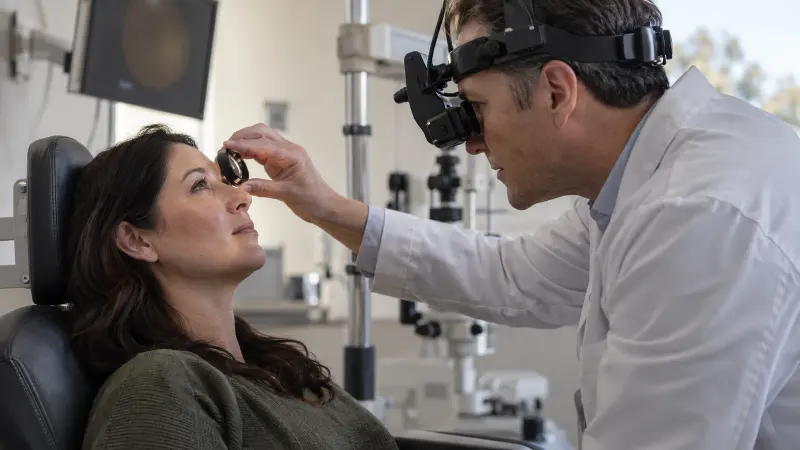
During an eyelid lesion visit, the clinician may flip the lid, check the tear film, examine the lashes, and compare both eyelids. If a lesion is photographed, the image helps show whether it is changing over time. If removal or biopsy is recommended, ask what tissue will be tested and what follow-up is needed.
- Tell the doctor when the lesion first appeared.
- Share any history of skin cancer or heavy sun exposure.
- Bring a list of eye makeup, lash products, and drops used near the lid.
- Ask whether warm compresses are appropriate or whether they delay needed care.
Small eyelid procedures can make people nervous because the area is close to the eye. Asking about numbing, healing, and warning signs can make the plan clearer.