An OCT scan in glaucoma uses light to create cross-sectional images of the optic nerve and retina. It measures layers that contain nerve fibers and ganglion cells, then compares those measurements with prior scans and a reference database. The scan can support diagnosis and monitoring, but it cannot confirm glaucoma by itself.
At a Glance
What patients should know
- OCT is quick, noninvasive, and usually does not touch the eye.
- The scan measures structure rather than side vision.
- Trend over time can matter more than one color-coded result.
- Eye pressure, visual fields, and the optic nerve exam remain essential.
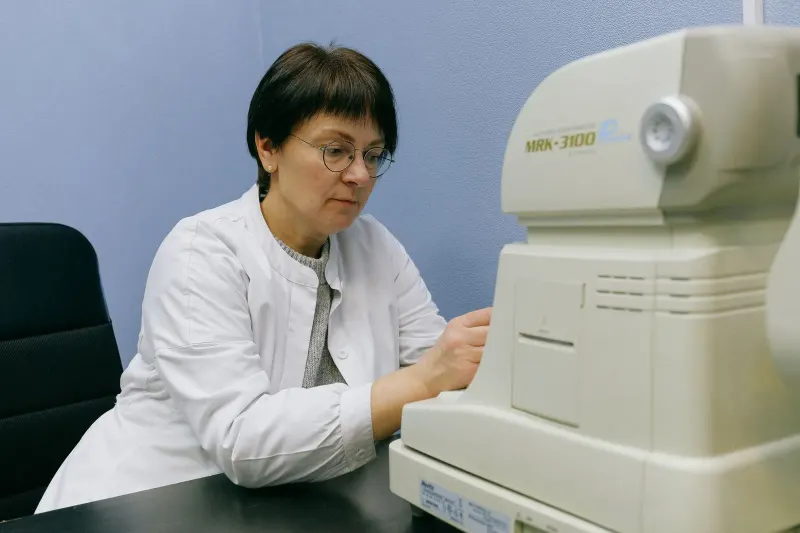
A picture and a measurement
Optical coherence tomography sends light into the eye and measures how it reflects from tissue. The American Academy of Ophthalmology EyeWiki overview explains that OCT can measure the retinal nerve fiber and ganglion cell layers. These structures can thin when glaucoma damages the optic nerve.
Which parts of the eye an OCT scan measures
The retinal nerve fiber layer
Nerve fibers travel across the retina and gather at the optic nerve. The scan measures their thickness around the nerve head. A thin area may support concern for glaucoma, but natural anatomy, high nearsightedness, prior optic nerve injury, and scan quality can also change the result.
The ganglion cell layer
Ganglion cells help carry visual information from the retina toward the brain. A macular scan can measure the region where many of these cells sit. Doctors compare the pattern with the nerve scan because the two views can provide complementary information.
The optic nerve head
Some reports show the shape of the nerve, its central cup, and the tissue around it. Cup size alone does not diagnose glaucoma. A large healthy nerve can have a large cup, while a smaller nerve can show damage with a less dramatic appearance.
How to read the color map without panicking
Green does not guarantee normal
Many reports compare your measurement with a group of people in a built-in database. Green often means the value falls within the expected range for that database. Early damage may still be present, especially if your prior scan was thicker or if the damaged region is small.
Red does not automatically mean glaucoma
Red or yellow can appear when anatomy differs from the reference group. High myopia, tilted nerves, segmentation errors, and other optic nerve conditions can cause a false alarm. The doctor should inspect the image, not just the summary colors.
Image quality changes confidence
Blinking, dry eye, cataract, poor focus, eye movement, and an off-center scan can reduce reliability. The technician may repeat the image during the same visit. Repeating a weak scan protects the quality of future comparisons.
Why doctors repeat OCT scans
A baseline creates a reference
The first good scan shows where your measurements started. A second scan may be obtained soon afterward to confirm repeatability. Later scans can then be compared with that baseline instead of relying only on an age-matched database.
Trend analysis looks for consistent loss
Software can plot thickness across several visits. Doctors look for a rate and pattern of change that repeats in the same area. One lower value may reflect measurement noise, while a consistent decline can support a treatment discussion.
Advanced disease can reach a measurement floor
When nerve layers become very thin, OCT may have less ability to show additional loss. Visual fields and the clinical exam become even more important. A stable thickness number does not always mean advanced glaucoma has stopped changing.
How an OCT scan in glaucoma fits with other tests
Visual fields measure function
OCT shows structure. A visual field test checks how well you detect lights in central and side vision. Structural and functional results may change at different times, so doctors use both rather than choosing one as the final answer.
Eye pressure shows one risk factor
Pressure can vary during the day and does not show the amount of nerve damage. Some people develop glaucoma at pressures within the usual range, while others have higher pressure without detectable damage. The article on why glaucoma pressure is only part of the story explains this distinction.
The optic nerve exam adds clinical context
Dilation lets the doctor inspect the nerve and retina directly. Corneal thickness, drainage angle, family history, age, and other health factors also shape risk. The National Eye Institute glaucoma guide emphasizes regular comprehensive exams because early glaucoma often has no symptoms.
Preparing for an OCT appointment
Bring prior scans when changing clinics
- Ask the former clinic whether images and reports can be transferred.
- Bring your glaucoma medicine list and note missed doses.
- Tell the technician about severe dry eye or trouble holding still.
- Ask whether today's scan is being used as a new baseline.
Ask about the trend, not only the color
Useful questions include whether the scan quality was good, whether change repeats in the same region, and whether the visual field agrees. Ask how the result affects the monitoring interval or treatment goal.
Use the same testing conditions when possible
Comparison is strongest when later scans are centered well and interpreted with the earlier images. Tell the technician if the target is hard to see or your eye is watering. A repeat image taken immediately is more useful than quietly accepting a scan affected by movement, a blink, or poor focus.
Common Questions About OCT and Glaucoma
Does an OCT scan hurt?
The scan usually does not touch the eye. You look at a target while the device captures images with light.
Do I need dilation for OCT?
Many scans can be obtained without dilation, but small pupils, cataract, or the need for a full eye exam may lead the doctor to dilate your eyes.
Can OCT find glaucoma before a visual field test?
Structural changes can appear before a repeatable field defect in some eyes. The reverse can also occur, so both tests remain useful.
What if my scan is red but my pressure is normal?
The doctor should review anatomy, image quality, prior scans, visual fields, and other causes of nerve thinning. Normal pressure does not settle the diagnosis.