Retinal Tear Diagnosis at a Glance
What the exam will tell you, and what it usually feels like
A possible retinal tear is checked with a careful dilated examination of the retina. The pupil is widened with drops so the eye doctor can inspect the back of the eye, including the far edge of the retina where a tear may be located. 1 2 The visit is meant to answer four practical questions: Is there a tear or hole? Has any retina detached? Was the full retina visible? What needs to happen next?
You may be worried that the examination will hurt or that finding a tear will always mean surgery. The dilated examination is used to find a small retinal tear or detachment. It is usually painless. Pressure used to inspect the retinal edge may feel uncomfortable, and the drops can cause blur and light sensitivity for a few hours. 1 3 If a tear is found, the next step may be laser, freezing treatment, or observation for some low-risk tears without symptoms. 2
Why the examination matters even when vision seems clear
A retinal tear may cause new floaters or flashes, but some tears cause no noticeable symptoms. A shadow or curtain and blurred vision can occur when bleeding or retinal detachment is also present. 2 Because these symptoms overlap with posterior vitreous detachment, the eye examination is what separates the findings. 4
What the Doctor Is Looking For
The doctor is checking for several different findings
A retinal tear forms when the vitreous gel pulls on the retina. A retinal hole develops from progressive thinning and is often smaller and lower risk, although some holes are treated like tears. 2 A retinal detachment means the retina has pulled away from its normal position at the back of the eye. The examination is used to identify which finding, if any, is present. 1 2 The clinician should tell you which term describes your result rather than using them as if they mean the same thing.
Posterior vitreous detachment is another possible result
Posterior vitreous detachment, often shortened to PVD, means the vitreous has separated from the retina. It is common for this separation to happen without a tear, but traction at a firmly attached area can tear the retina and may lead to retinal detachment. 4 2 This is why the result conversation should cover both the vitreous and the retina.
How to Prepare for the Retinal Examination
Describe the change before the drops go in
Tell the clinic when the symptom began, which eye seems affected, whether it is changing, and whether you see flashes, new floaters, blur, or a missing area. Mention a recent eye injury, earlier retinal tear or detachment, and previous eye surgery. If you cannot tell which eye is affected, say so. Bring your current medicine list and the name of any eye drops you use. For a related symptom pattern, read What a Weiss Ring Floater Means for the Vitreous and Retina.
Plan for blur and light sensitivity afterward
For a few hours after a dilated eye examination, vision may be blurry and the eyes may be sensitive to light. The National Eye Institute recommends asking a friend or family member to drive you home and bringing sunglasses for comfort. 3 Make the transport plan before the visit if possible. Ask the clinic about its driving advice if you are uncertain.
Bring the questions that will close the loop
Write down what you need to know: whether the entire peripheral retina was visible, whether a tear, hole, PVD, bleeding, or detachment was found, and when the next examination is due. Bring contact details for another eye clinic if records need to be shared.
What Happens During the Examination
Vision and eye checks may come first
A dilated eye visit may begin with checks of visual clarity, side vision, eye movements, pupil responses, and eye pressure. 3 The checks do not all have to occur in the same order.
Dilating drops widen the pupil
Dilating drops widen the pupil so more of the inside of the eye can be seen. Dilation is part of creating the view for the examination; it is not a diagnosis by itself. 3 The staff may ask you to wait while the drops take effect. Tell them about discomfort, questions, or any difficulty holding your eye open. Ask for a brief pause when the clinician changes position.
Indirect ophthalmoscopy gives a wide retinal view
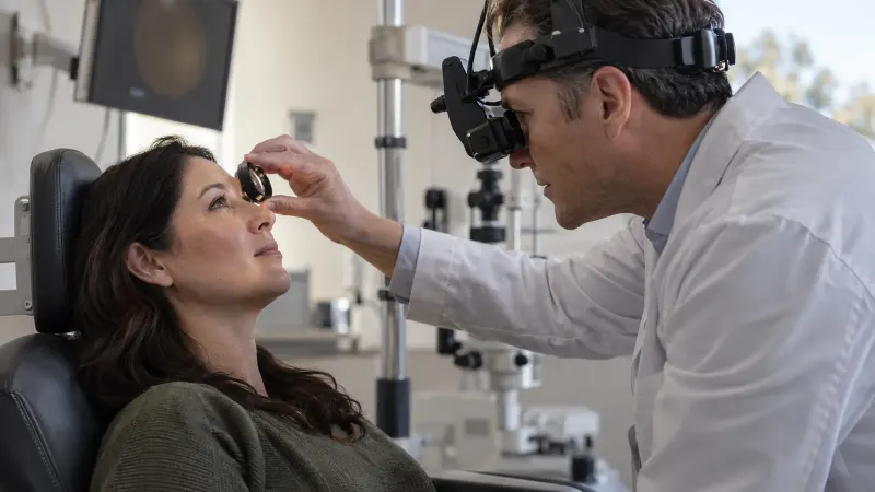
Indirect ophthalmoscopy with scleral indentation is a preferred technique to confirm PVD and exclude retinal tears or retinal detachment. 4 Follow the examiner’s instructions about where to look.
Scleral depression helps expose the far periphery
During scleral depression, the examiner applies slight pressure at the outside of the eye or over the eyelid while viewing the retina. This helps inspect the peripheral retina and can be uncomfortable for some people. 2 1 Tell the clinician if you need a pause.
A three-mirror lens is another examination method
A slit-lamp examination with a three-mirror contact lens is another method used to look for retinal tears or detachment. 4 2 The clinician chooses the method that provides the needed view. Ask which method was used if you want it recorded for follow-up.
How to Understand the Result
If a retinal tear is found, the next step is not always the same
A retinal tear is not the same finding as a retinal detachment. Retinal tears are often treated with laser or freezing treatment, but not every retinal tear requires treatment. Some low-risk tears without symptoms can be observed. 2 1 The clinician will tell you whether the finding is a tear or a hole and whether retinal detachment is also present. Ask why treatment or observation is recommended for this finding and when the next check is due.
If retinal detachment is found
Treatment for retinal detachment depends on its type and extent. A small hole or tear may be sealed with laser or freezing treatment, while a larger detachment may require surgery to move the retina back into place. Prompt treatment can help protect vision. 1 Ask who will provide the next assessment, where it will happen, and how soon you should go.
If no tear is seen in a complete view
After an acute PVD, another dilated retinal examination may be planned because a new retinal break or detachment can appear while the PVD is evolving. “No tear seen today” should come with a follow-up plan. 4 Ask when to return and which symptom changes should prompt an earlier call.
If blood or another opacity blocks the view
When hemorrhage or another opacity prevents a complete retinal view, ocular ultrasound may help detect retinal detachment or a large flap tear. Ultrasound cannot rule out every retinal tear. 4 A meta-analysis found emergency-practitioner ultrasound accurate for retinal detachment, while evidence for retinal tears showed poor sensitivity; timely ophthalmology follow-up is still needed when a tear remains possible. 5 Ask what part of the retina remained unseen and how the gap will be closed.
Discomfort, Limitations, and Follow-Up
Most discomfort is brief
The dilated retinal examination is usually painless, although pressure on the eyelids during the search for a tear may feel uncomfortable. Blur and light sensitivity may continue for a few hours after dilation. 1 3 Tell the team if the examination is difficult so they can guide you through each step.
One test may not answer every question
A careful peripheral retinal examination is the main step in retinal tear diagnosis. Ultrasound is useful when the view is blocked, but a negative ultrasound does not exclude all tears. 2 4 Separate new tears can develop after an earlier tear has been treated, so continued monitoring may still matter. 2 The useful outcome is a clear result plus a plan for any remaining uncertainty.
When to Seek Care Before or After the Test
Get immediate eye care for detachment warning signs
Go to an eye doctor or emergency department right away for a sudden increase in floaters, flashes of light, or a dark curtain or shadow over part of the vision. Retinal detachment is an emergency, and prompt treatment can help protect vision. Do not wait for a routine appointment if these symptoms are new. 1 5 These symptoms do not confirm a detachment by themselves; a dilated retinal examination is needed to identify the finding. 4 1
Report a clear change after a reassuring examination
After a PVD assessment, new or increasing flashes or floaters and a new loss of peripheral vision need prompt reassessment because a retinal break or detachment can develop later. 4 When you call, state what changed and when, which eye is affected, and what the last examination found.
Keep follow-up when the view was incomplete
If bleeding or another opacity blocked the retinal view, a negative ultrasound may still need close ophthalmology follow-up because ultrasound is less reliable for retinal tears than for retinal detachment. Keep the arranged repeat examination unless the treating clinician changes the plan. 5
Questions About the Examination
Is a retinal tear examination painful?
The dilated examination is usually painless. The part that may feel uncomfortable is pressure on the eyelid or outside of the eye while the clinician checks for peripheral tears. 1 Tell the examiner if you need a pause or if the light is hard to tolerate.
Why do my pupils need to be dilated?
Dilating drops widen the pupil and allow the eye doctor to see more of the inside of the eye. A dilated examination can reveal a small retinal tear or detachment before it affects vision. Dilation creates the view needed for the clinician to inspect the retina. 3 1
Can I drive myself home after dilation?
The National Eye Institute advises asking a friend or family member to drive you home because dilation can cause temporary blur and light sensitivity. 3 Plan transport before the appointment rather than deciding after the drops are given. If your clinic gives different instructions for your situation, follow those instructions.
What is scleral depression?
Scleral depression is a technique that applies slight pressure to the eye while the clinician examines the peripheral retina. Retina specialists use it, or a three-mirror lens, as an important part of looking for a retinal tear. You may feel pressure during this part of the examination. 2 Ask for a pause if you need one.
Can a retinal photograph replace the examination?
The preferred methods for excluding a retinal tear are indirect ophthalmoscopy with scleral indentation or slit-lamp examination with a three-mirror lens. 4 Ask whether the far peripheral retina was directly examined and whether any area could not be seen well.
Why might the doctor order an ultrasound?
Ocular ultrasound can help when blood or another opacity blocks the view of the retina. It is useful for detecting retinal detachment and some large tears, but it cannot rule out every retinal tear. 4 5 The follow-up question is what the ultrasound showed and whether another dilated examination is needed when the view clears.
Questions About Results and Next Steps
Does “no tear seen” mean I need no follow-up?
Not always. After an acute PVD, clinicians often repeat the dilated examination because a new tear or detachment can develop while the vitreous separation is evolving. Use the return instructions sooner if symptoms change. 4 Ask whether the first examination gave a complete view and whether your specific result needs another appointment.
Can ultrasound alone rule out a retinal tear?
No. Ultrasound can be accurate for retinal detachment, but it is less sensitive for retinal tears. A negative ultrasound should not replace timely ophthalmology follow-up when a tear is still possible. 5 4 Ask what the ultrasound was designed to detect and how the clinician plans to examine any retina that was hidden.
Does every retinal tear need laser treatment?
No. Many retinal tears are treated with laser or freezing treatment, but some low-risk tears without symptoms can be observed. The decision depends on the actual finding. 2 Ask the specialist why treatment or observation fits your tear and what follow-up is required. Do not use another person’s result to choose your plan.
How long will my vision stay blurry after dilation?
Blur and light sensitivity may last for a few hours after a dilated eye examination. 3 The exact experience varies, so use the clinic’s instructions for its drops. Arrange a ride home before the visit if needed, and bring sunglasses if they help. Contact the clinic if you have a concern after the examination.
Questions to Ask Your Doctor
- Did you see the entire peripheral retina?
- Was the result a PVD, retinal hole, retinal tear, or retinal detachment?
- If no tear was seen, when should the retina be examined again?
- If the view was limited, what will close the gap?
- Which symptoms mean I should return immediately?
- Who should I call outside office hours?
Sources
- National Eye Institute (2025). Retinal Detachment.
- American Society of Retina Specialists (2026). Retinal Tears.
- National Eye Institute (2025). Get a Dilated Eye Exam.
- American Academy of Ophthalmology EyeWiki (2026). Posterior Vitreous Detachment.
- JAMA Network Open (2020). Ocular Point-of-Care Ultrasonography to Diagnose Posterior Chamber Abnormalities: A Systematic Review and Meta-analysis.