Anterior Uveitis Diagnosis at a Glance
Will the visit hurt?
Short answer: A dilated eye exam is simple and painless. It can include checks of sight, pupil response, and eye pressure.1 Uveitis can cause a painful, red eye that is sensitive to light.2 Tell the examiner if pain or light makes a step hard, and ask for a pause.
The drops may blur sight and make the eyes sensitive to light for a few hours.1 Ask about driving before the visit. Arrange a ride if you do not feel safe traveling after dilation.
Symptoms may point to anterior uveitis. To confirm it, the clinician must see cells in the front chamber with a slit lamp.3
The visit answers more than one question
The clinician uses the location, duration, appearance, laterality, signs, and symptoms to characterize the uveitis and narrow possible causes.4
Anterior uveitis is classified by features such as onset, duration, whether one or both eyes are involved, and whether the inflammation is limited to the front of the eye.3 Ask how those features affect the questions, tests, and follow-up plan in your case.
What the Clinician Is Looking For
Inflammatory cells and protein in the front chamber
Inflammatory cells and protein in the front-chamber fluid appear as cells and flare during examination; anterior-chamber cells are diagnostic of anterior uveitis, while flare alone does not necessarily mean inflammation is active.4
Slit-lamp assessment can also show deposits on the back of the cornea, changes in the iris or pupil, and adhesions between the iris and the lens.4 Ask which signs were present and how they will be compared at later examinations.
Vision, eye pressure, and the rest of the eye
A dilated eye examination includes visual-acuity, pupil-response, and eye-pressure tests, followed by dilation so the clinician can see inside the eye.1
People with acute anterior uveitis should initially have dilation so the clinician can examine the fundus and check whether inflammation also involves the back of the eye.3
Other conditions that can resemble uveitis
AAO EyeWiki notes that the ophthalmologist must distinguish acute anterior uveitis from inflammation or infection in other eye structures, including conjunctivitis, keratitis, and scleritis.3 Ask which similar conditions were considered during the examination.
How to Prepare for the Diagnostic Visit
Bring the timeline and any prior eye records
Write down when symptoms began, whether they started suddenly or gradually, and whether they have changed. Note which eye is affected and whether a similar episode happened before. Bring records from prior episodes if you have them, including the diagnosis, examination findings, tests, medicines used, and response. For a related symptom pattern, read Anterior Uveitis Symptoms to Take Seriously.
Whether symptoms began suddenly or gradually, involve one or both eyes, and fully resolve or recur are features used to classify uveitis.3 Use the short written timeline during the examination.
List medicines, illnesses, and symptoms elsewhere
Bring a current list of prescription medicines, nonprescription products, injections, and eye drops. Mention recent eye surgery or injury. Tell the clinician about known infections, immune conditions, and any recent illness or travel that seems relevant. You can compare this topic with Urgent Symptoms of Anterior Uveitis.
The diagnostic history may include questions about joint or back symptoms, skin changes, bowel symptoms, mouth or genital ulcers, breathing symptoms, and past infections because some uveitis patterns are associated with disease outside the eye.4 Answer what you know without trying to select a diagnosis yourself.
Plan for dilation and temporary blur
A dilated eye examination is generally simple and painless, but the drops can cause temporary blur and light sensitivity for a few hours.1 Bring sunglasses if they help. Ask the clinic before the visit whether you should arrange a ride, especially if you have not been dilated before or do not feel safe driving with blurred vision.
Tell the clinician about a previous reaction to eye drops. Bring the glasses or contact lenses you normally use, but follow the clinic’s instructions about whether to remove contact lenses before the examination. For another care decision in this area, see When Intermediate Uveitis Symptoms Are Urgent.
What Happens During the Examination
Vision and pupil checks come first
Visual-acuity testing checks how clearly you see, and pupil-response testing checks how the pupils react when light enters the eyes.1
Tell the examiner if you cannot keep the eye open because of pain or light sensitivity. Ask for a pause if you need one.
The slit-lamp examination uses a bright focused light
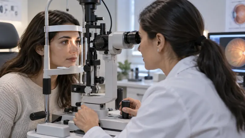
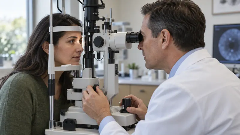
The slit lamp is the main examination tool for acute anterior uveitis because it can detect white blood cells in the fluid at the front of the eye.3 Keep your chin and forehead in position as directed, and tell the examiner if you need a pause.
Clinical characterization at the slit lamp helps determine whether the inflammation is granulomatous or non-granulomatous and whether findings suggest a particular pattern or cause.4 Ask the clinician to explain these appearance terms in plain language.
Pressure and dilation add information
Tonometry measures eye pressure with a quick puff of air or gentle contact with a special tool, and dilating drops widen the pupil so the clinician can see inside the eye.1
The National Eye Institute lists a dilated eye examination and medical history as central parts of checking for uveitis and notes that additional tests may be recommended when infection or another disease is suspected.5 Ask which additional test, if any, is recommended for your findings.
How Results Guide Additional Testing
The eye pattern narrows the possibilities
Characterizing uveitis by location, course, appearance, laterality, and clinical signs helps the clinician form a working diagnosis and target the workup.4
Laboratory testing for acute anterior uveitis is applied selectively according to the clinical pattern and suspected cause; broad, non-targeted survey testing is discouraged because it has low predictive value and rarely changes management.3 Ask what question each proposed test is meant to answer.
Blood tests and imaging are not the same for everyone
AAO EyeWiki describes testing for HLA-B27 and syphilis in the basic acute-anterior-uveitis workup, chest imaging when bilateral granulomatous disease raises concern for sarcoidosis, and other laboratory tests based on clinical suspicion.3 Ask what question each proposed test or image is meant to answer.
The randomized ULISSE trial included 903 people with uveitis; in its standardized group, further investigations could be performed according to ophthalmological findings.6 Ask how the study's approach relates to your individual findings.
Sometimes no underlying cause is identified
Doctors do not always identify the cause of uveitis, even though inflammation is present.5 If the clinician uses the word “idiopathic,” ask what was evaluated and what the term means in your case.
Ask what would make the team revisit the cause: recurrence, involvement of the other eye, new symptoms elsewhere, a different slit-lamp pattern, or lack of the expected response. Keep a copy of test results so future clinicians can see what has already been checked.
What the Diagnosis May Show Next
Confirmed inflammation limited to the front of the eye
Anterior-chamber cells confirm anterior uveitis, while a dilated fundus examination helps determine whether there are signs of intermediate or posterior involvement.4
Anterior uveitis may be acute, recurrent, or chronic, and the course helps shape the investigation and follow-up.3 Ask the clinician to write down which type they believe is present and what findings support it.
A complication or inflammation beyond the front chamber
Anterior uveitis and its treatment can be associated with complications including raised eye pressure, glaucoma, cataract, and macular edema.4 Ask what any identified complication risk means for you and what will be checked at follow-up.
A different cause of the red or painful eye
When the required anterior-chamber cells are absent, the clinician considers other diagnoses; conjunctivitis, keratitis, and scleritis are among the conditions that can resemble acute anterior uveitis.3
Ask what was ruled in, what was ruled out, and which change should prompt another examination. If the diagnosis remains uncertain, ask when and where it should be rechecked rather than assuming that all red-eye conditions follow the same course.
When to Seek Prompt Eye Care
Get prompt assessment for a painful red light-sensitive eye
Acute anterior uveitis can cause a very painful red eye, sensitivity to light, and reduced vision, and the National Eye Institute advises seeing an eye doctor right away when uveitis symptoms occur because untreated inflammation can cause vision loss.25 Use the prompt eye-care route available in your area rather than waiting for a routine appointment.
If you cannot reach the usual clinic, use the urgent route available in your area. Tell the service when symptoms began and whether vision has changed. Because acute anterior uveitis requires inflammatory cells to be detected in the front chamber, a photograph or message cannot confirm the diagnosis.3
Call the same day if symptoms worsen before review
If eye pain, light sensitivity, redness, or blurry vision appears or worsens, seek prompt eye care because untreated uveitis can cause vision loss.5 Follow any emergency instructions the clinical team has already given you. If symptoms follow an eye injury or surgery, state that clearly when you call.
Tell the clinician if an earlier episode felt similar, but arrange the recommended examination for the current symptoms.
Keep planned follow-up even when the eye feels better
At follow-up, ask the clinician to compare inflammatory cells, vision, eye pressure, and any complications with the prior examination. Bring your medicines and the written schedule so the team knows exactly what you used.
If transport or cost may prevent attendance, tell the clinic before the visit. Ask which part of the follow-up is time sensitive and whether a closer qualified eye service can perform it.
Questions About the Examination
Can a regular photo diagnose anterior uveitis?
No. A regular photograph cannot confirm acute anterior uveitis because diagnosis requires inflammatory cells in the front chamber to be detected with a slit lamp.3 Follow the recommended plan for an in-person eye examination.
Is the slit-lamp examination painful?
The National Eye Institute describes a dilated eye examination as simple and painless and says pressure testing may use a quick puff of air or gentle contact with a special tool without hurting.1 Anterior uveitis itself can make the eye sensitive to light.2 Tell the examiner if the light is uncomfortable or you need a pause. Ask the clinician to explain each step before it happens.
Will my eyes be dilated?
People with acute anterior uveitis should initially undergo dilation so the examiner can check the fundus, and the drops can cause temporary blur and light sensitivity for a few hours.31 Ask about driving before the appointment and arrange a ride if you do not feel safe traveling home with dilated vision.
Why is eye pressure measured?
A rise in intraocular pressure and subsequent glaucoma are recognized complications of acute anterior uveitis.3 Ask how the pressure result compares with prior measurements and whether it changes the plan.
Does every patient need blood tests?
No single broad test panel fits every case. Current AAO EyeWiki guidance discourages a non-targeted uveitis survey and recommends testing guided by the clinical pattern and suspected cause.3 Ask what each test could find and how a positive or negative result would change the plan.
Questions About Results and Follow-Up
What does “cells and flare” mean?
Cells are inflammatory cells in the front-chamber fluid, while flare comes from protein in that fluid; cells are diagnostic of anterior uveitis, but flare alone does not necessarily show active inflammation.4 Ask how the finding was graded and whether other examination findings change its meaning.
What if all of my laboratory tests are negative?
The diagnosis of anterior uveitis rests on inflammatory cells seen in the front chamber, while doctors do not always identify an underlying cause.35 Ask what was tested, which causes were considered, and what later changes would lead the team to repeat or expand the investigation.
Will I need to see another specialist?
Ask the treating clinician whether another specialist is recommended, why the referral is being made, and how soon the visit should occur. Bring the eye-clinic records, test results, medicine list, and a short symptom timeline to the referral.
Can the diagnosis change at a later visit?
Yes. Clinical reviews describe uveitis diagnosis as a process guided by the pattern and evolution of findings, with targeted investigations added when appropriate.4 Ask which new finding could make the working diagnosis more specific, and keep prior records so the next clinician can compare episodes.
Questions to Ask Your Doctor
- What findings confirm anterior uveitis in my eye?
- Is inflammation limited to the front of the eye?
- Are my vision or eye pressure affected?
- What cause does the pattern suggest, if any?
- What is each proposed blood test or scan meant to answer?
- What changes should make me call the same day?
- When and where should the eye be examined again?
Sources
- National Eye Institute (2025). Get a Dilated Eye Exam.
- BMJ Clinical Evidence (2010). Uveitis (acute anterior).
- American Academy of Ophthalmology EyeWiki (2026). Acute Anterior Uveitis.
- Clinical Optometry (2016). Diagnosis and treatment of anterior uveitis: optometric management.
- National Eye Institute (2024). Uveitis.
- PLOS ONE (2020). Evaluating the cost-consequence of a standardized strategy for the etiological diagnosis of uveitis (ULISSE study).