Meibography for dry eye shows the structure of the meibomian glands, which sit inside the eyelids and help make the oily layer of the tear film. When these glands are blocked, shortened, or lost, tears may evaporate too quickly.
Dry eye is not always from too few tears. Many people have evaporative dry eye, meibomian gland dysfunction, or a mix of tear film problems. Meibography can help explain why symptoms persist even when the eye looks only mildly red.
At a Glance
- Meibography images the oil glands inside the eyelids.
- The test can show gland shortening, dropout, or uneven structure.
- Results are interpreted with symptoms, tear film testing, lid findings, and medical history.
- A painful red eye with reduced vision, contact lens pain, or a white corneal spot needs prompt care.
Why Meibography For Dry Eye Matters
The tear film has layers, and the oily layer slows evaporation. If meibomian glands are not working well, the eyes may burn, water, fluctuate in clarity, or feel gritty, especially during screen use or windy conditions.
Seeing the glands can motivate a more tailored plan. It may also help set expectations. Gland structure can sometimes look permanently changed, so the goal may be symptom control and preventing worsening rather than restoring every gland.
Dry eye organizations and ophthalmology guidance increasingly distinguish evaporative dry eye from aqueous tear deficiency. Meibography is useful because it shows a structure that cannot be judged fully from symptoms alone.
What the Test Looks For
During meibography, the eye care team is looking for patterns that match the symptoms and the medical question. The details matter because similar complaints can come from different parts of the visual system.
The result is most useful when it is repeatable and when it fits the rest of the examination. If the result does not fit, repeating the test or using a different method can be the careful choice.
For meibography for dry eye, patients should describe what has changed in ordinary life, not only what happens in the exam room. Reading, driving, screen use, sports, glare, balance, pain, or one-eye differences can make the result more meaningful for dry eye disease care.
- Whether meibomian glands are visible, shortened, or missing
- Whether gland loss is worse in the upper or lower lids
- Whether symptoms match lid margin inflammation or poor oil flow
- Whether treatment goals should focus on oil quality, tear stability, or inflammation
What Results Can and Cannot Tell You
Meibography is not a complete dry eye workup by itself. Tear breakup time, staining, lid evaluation, symptoms, medications, autoimmune history, and environment can all matter.
A dramatic image does not always equal severe symptoms, and mild images do not always mean mild discomfort. The clinician should interpret the scan with the full exam.
The scan also cannot prove which treatment will work best. It helps guide the conversation, but response varies with inflammation, habits, skin conditions, contact lens wear, and other health factors.
- Ask what the result means for your specific diagnosis.
- Ask whether the finding is new, stable, or uncertain.
- Ask whether repeat testing or imaging is recommended.
- Ask what symptoms should prompt faster contact before the next visit.
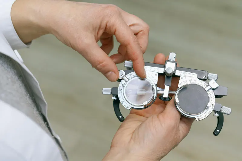
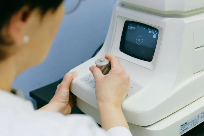
What to Expect at the Appointment
During meibography, the eyelid is usually gently positioned so the device can image the glands. The test is often quick, but patients should speak up if lid handling is uncomfortable.
The clinician may also press lightly on the lid margin to see whether oil flows easily and whether it is clear, cloudy, thick, or absent. That functional finding can be as important as the image.
Afterward, the discussion should connect the scan to practical goals such as more stable vision, less burning, better contact lens tolerance, or fewer flare-ups.
- Tell the clinician about screen time, contact lens wear, and prior eyelid treatments.
- List medications that may worsen dryness.
- Mention rosacea, autoimmune disease, allergies, or hormone changes.
- Ask what findings suggest inflammation, gland blockage, or aqueous tear deficiency.
When to Seek Faster Eye Care
Dry eye is usually chronic, but severe pain, marked light sensitivity, a red eye with reduced vision, contact lens-related pain, or a white spot on the cornea needs prompt evaluation.
If symptoms are severe, sudden, or clearly different from your usual pattern, it is safer to ask for guidance promptly. Routine testing is valuable, but urgent symptoms need timely examination.
How Follow-Up Uses the Findings
Follow-up for dry eye disease often depends on whether results are stable over time. One visit may set a baseline, while later visits show whether vision, eye structure, or symptoms are changing.
Patients can help by keeping appointments, reporting changes early, and bringing questions about how the result affects daily activities. The best plan connects test results with the person, not just the printout.
It is also fair to ask how meibography will change decisions today. Sometimes the answer is treatment, but often it is a cleaner baseline, a safer monitoring interval, a referral, or a repeat test under better conditions. That context keeps the visit from feeling like a pass-fail exercise and makes the next step easier to understand.
If the finding affects work, school, sports, reading, driving, or home safety, say that clearly. Functional details help the clinician connect meibography for dry eye results with practical advice and realistic follow-up timing.
Common Patient Questions
Does gland loss grow back? Lost gland structure may not fully return, but symptoms can often be managed and remaining glands supported.
Is watering a dry eye symptom? Yes. Irritated eyes may reflexively water even when the tear film is unstable.
Do I need meibography at every visit? Not usually. It may be repeated when tracking change or planning treatment, depending on the clinician.